Scaffolds for Tissue Engineering

Autoclavable soft actuators made of medical silicone elastomer for adapted stimulation of cell growth

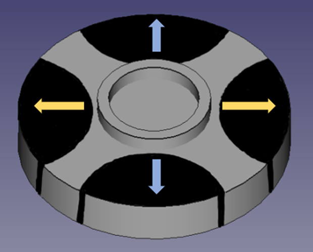
Dielectric elastomer actuator for applying biaxial stress on tissues
© Fraunhofer ISC
Dielectric elastomer actuator for applying biaxial stress on tissues placed in the circular well

CeSMA develops soft actuators made of medical grade silicone elastomer that are autoclavable and can therefore come into direct contact with biological cells.

The aim is to control and promote both cell growth and the direction of growth through biaxial, mechanical stimulation. By using electrically conductive silicone, the cells can be electrically stimulated at the same time, allowing them to differentiate as i.e. muscle cells. At moderate frequencies of around 1 Hz, the actuators can perform an actuation of up to 10 %. Cell adhesion is adjusted by surface pretreatment and structuring of the silicone film.
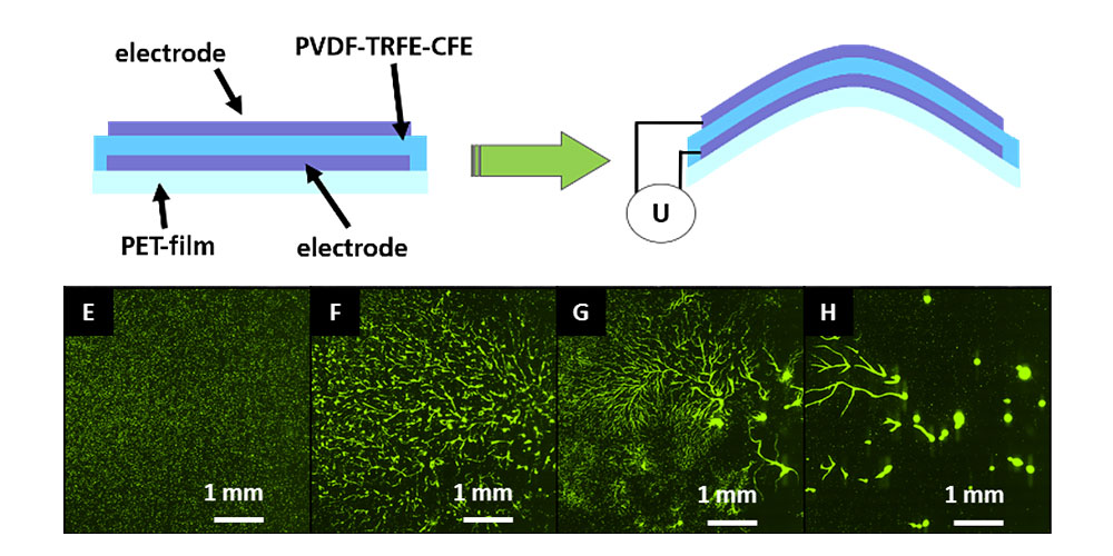
To stimulate and control cell growth, fields of microactuators (arrays) can also be produced. These can be made from piezoelectric polymers, which can achieve a displacement of up to 10 µm over a wide frequency range. Since these actuators also exhibit good transparency, cell growth can be easily monitored using optical methods (e.g., fluorescence microscopy). The arrays are produced using screen printing or inkjet printing.

Scaffolds for Tissue Engineering
© Fraunhofer ISC
Operation of piezoelectric actuation (top) and example measurements on keratinocytes under stimulation (bottom).

Customized 3D structures for cell cultivation and tissue maturation

Biocompatible ORMOCER® materials can be processed into virtually any 3D structure at the micrometer scale using two-photon polymerization (2PP). The sterilization of this material class in an autoclaving process allows the produced structures to come into direct contact with biological cells.

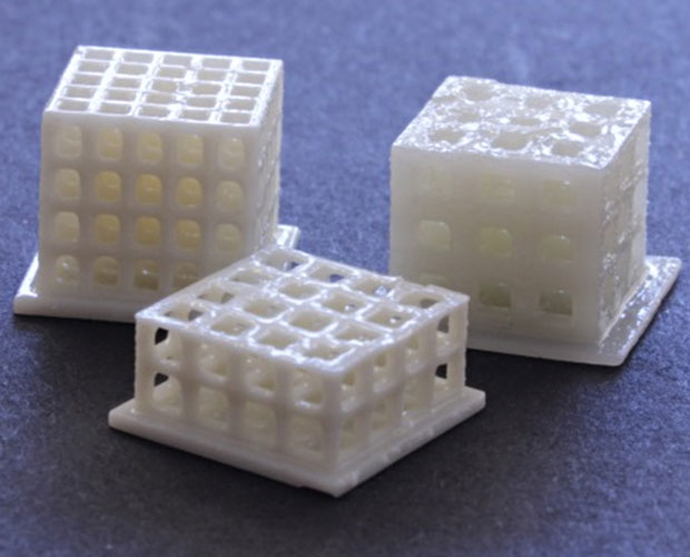
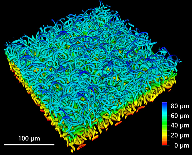
A fundamental approach in the field of biofabrication is to grow viable cells on a 3D scaffold that provides adhesion for further cell cultivation and tissue maturation. Since the cells can perceive this environment with submicrometer resolution and are influenced by the 2D and 3D morphology of the substrate, two-photon polymerization is ideally suited for generating such scaffold structures. Both solid three-dimensional templates and soft cell scaffold structures based on hydrogels, consisting of loosely interconnected rings with a diameter of 20 µm, can be created. These structures have been used to culture cardiomyocytes derived from hiPS.

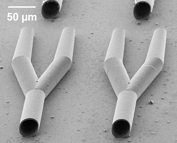
Non-biological vascular replacement structures can also be produced using this method, which can be equipped with additional surface structures or openings.

Biodegradable materials ORMOCER®
© Fraunhofer ISC
Three-dimensional template as scaffold for tissue engineering.
Hydrogel cell scaffold structure
© Fraunhofer ISC
Hydrogel cell scaffold structure made of loosely interconnected rings with a diameter of 20 µm.
2PP Microoptics CeSMA
© Fraunhofer ISC
Microvessels produced by 2PP in biocompatible ORMOCER®.
2PP structures ear canal prostheses
© Fraunhofer ISC
Structures produced by 2PP as prothesis in the ear canal.

HiPS derived cardiomyocite cells

Cultured on top of a 2PP structured hydrogel scaffold

Tissue engineering at the Fraunhofer Translational Center for Regenerative Therapies TLC-RT in Würzburg

Tissue engineering refers to the artificial production of biological tissue and is an important part of regenerative medicine. One area of tissue engineering is the development of in vitro test systems, which can be used for researching various scientific topics such as development, infection, tumor, and pathobiology, as well as for drug and toxicity studies.

The Fraunhofer Translational Center for Regenerative Therapies TLC-RT at Fraunhofer ISC develops not only new in vitro tissue models and test systems but also production processes and biological implants, including prototypes that have a vascularized structure.